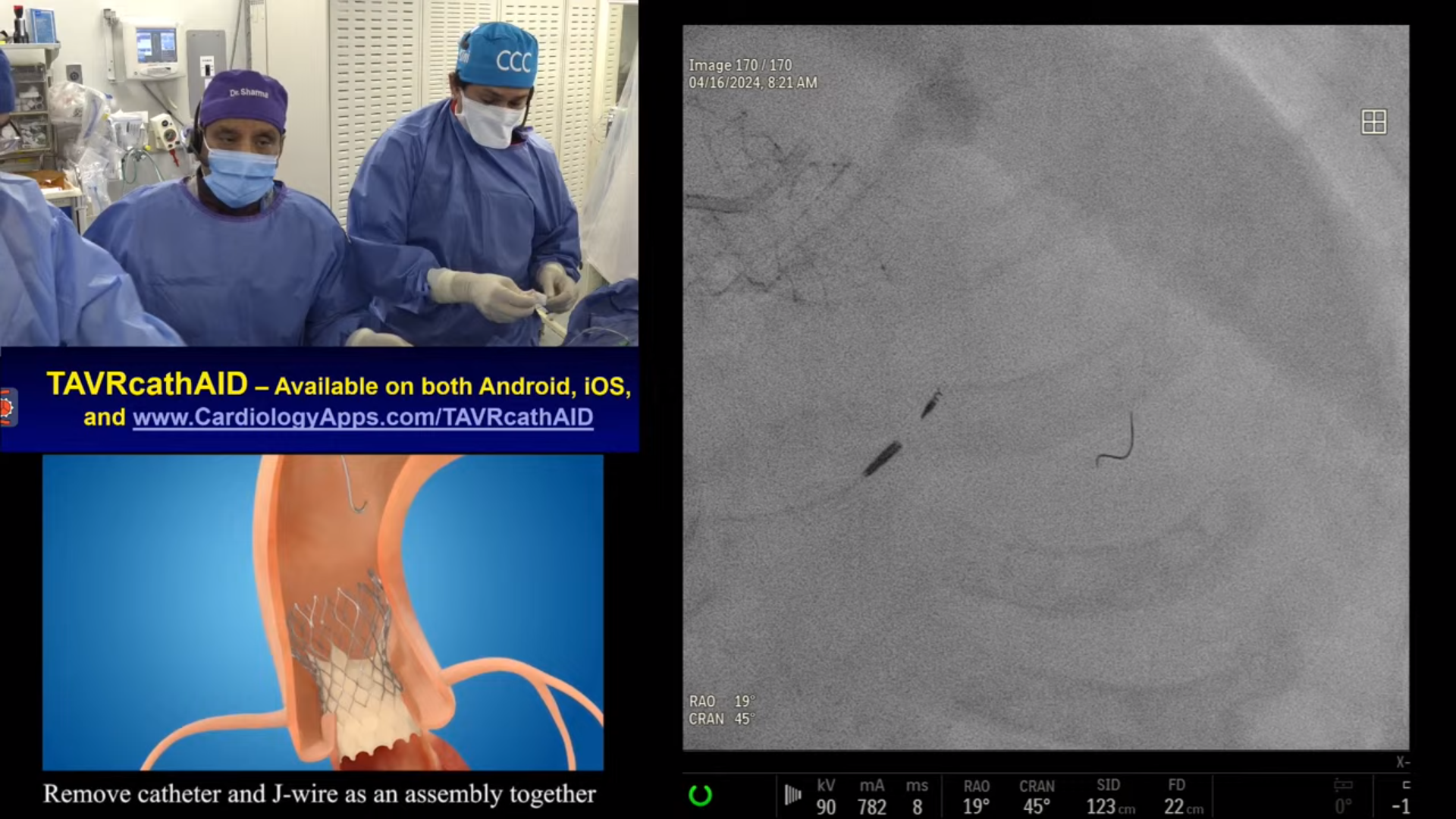
CASE & Plan:
76-year-old female presented with mild angina and a stress MPI as pre-op for shoulder surgery revealed multivessel inferior and lateral ischemia. A Cardiac Cath on July 31, 2023 revealed 2 V CAD: multiple calcified 80-95% lesions in RCA, 95% calcified angulated lesion in proximal LCx with SYNTAX Score of 20 and LVEF 50%. Patient underwent successful interventions of RCA using RA and 3 (Promus Elite) DES. Patient is now planned for staged PCI of angulated proximal LCx using Rotational Atherectomy and DES.
Q&A
Q
What are the most important considerations for selecting Rotablator (RA) for an angulated and calcified lesion?
A.
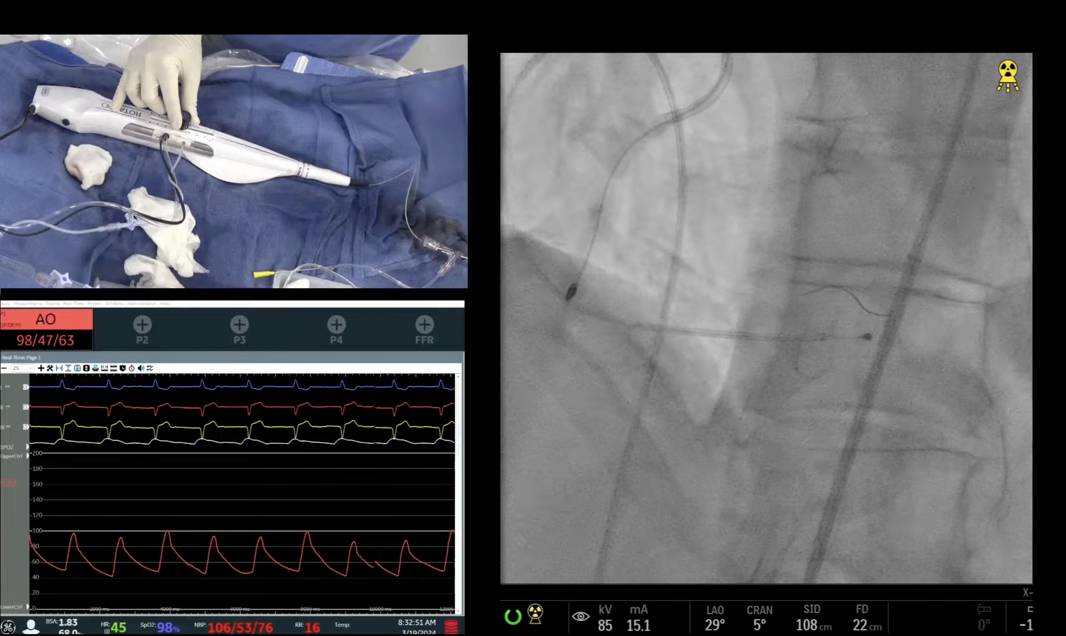
Most important and basic consideration, is to decide that do you absolutely need RA in these calcified angulated lesions, due to higher inherent complications of dissection and perforation; like our case where no other device could cross. If we can dilate the lesion by non-RA means like gradual POBA+/- IVL, then RA can be safely avoided. RA, certainly will have higher complications of dissection, wire bias and perforation in these lesions and we need to be prepared to tackle these procedural issues. Other procedural considerations will be to use the Rotafloppy wire, smallest burr possible, incomplete ablation of the lesion, lower RA speed of 120k and even a technique of bending the Rota wire to settle in the angulation.
Q
Why is Orbital Atherectomy (OA) not a good device for such lesions?
A.
The OA wire is 0.012”, stiffer compared to floppy 0.009”Rotawire and hence OA has more tendency to create dissections and wire bias in the angulated lesions. Therefore OA should be avoided in these super angulated lesion for concerns of potential dissection and perforation. Newer Viperwire Flextip is hydrophilic and is helpful in these lesions if OA is required. Disadvantage of Viperwire Flextip is that it provide less support then the conventional Viper wire.
Q
Should IVL be completely avoided for such lesions?
A.
IVL should be avoided as IVL balloon is stiff and will not make the turn.
Q
How would a cutting balloon perform in such cases?
A.
Cutting balloon may go after the RA or after other compliant ballon but should be used only after atherectomy; otherwise may create lesion closure or dissection if does not cross.
Q
The traditional teaching is to avoid laser in angulated lesions. Why did you use it?
A.
Same way laser catheter will not make the turn in the angulated lesions and may create dissection or perforation by preferentially delivering laser energy in the angulated eccentric lesions. Hence we did not use the Excimer laser in this case (although warmed up the machine) as Rota finally crossed at very slow speed of 120-130,000 rpm.
Q
If none of the ablation devices worked, would a high-pressure balloon do a sufficient job?
A.
I am skeptical that any balloon based technology would have worked in this severely calcified angulated lesion. If other devices could not cross then this pt needed to referred for elective in-hospital CABG.
Q
What were your considerations for selecting guidewires for such angulated lesions?
A.
Hydrophilic guidewires of Fielder, Whisper or Prowater type will be first line choice. If these guide wires could not cross despite using the angulated catheters, then stiffer wires like Pilot 50, MiracleBro 6 or Gaia 2 will be second line choice but little more risky as may go subintimal.
Q
Several operators would have jumped into using a guide extension catheter. Not a good idea?
A.
Guide extenders in this case after RA is appropriate to facilitate subsequent device passages delivery.
Q
What is the ideal stent for angulated lesions?
A.
Currently all stents are good for these angulated lesions with our preference being Synergy and Xience DES.
Q
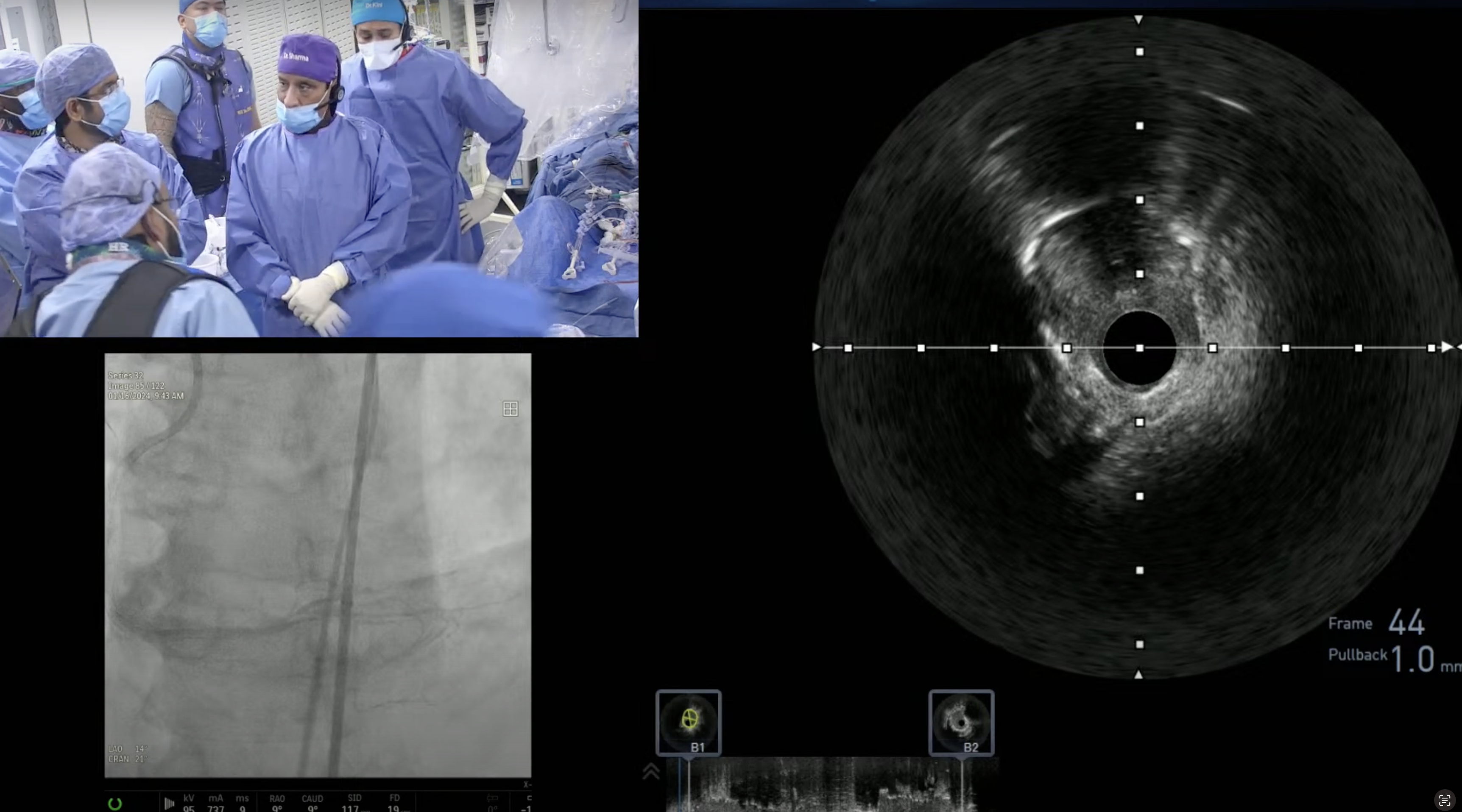
IVUS or OCT?
A.
IVUS will be preferred in these lesion over OCT because of its ability to cross angulated lesions while OCT being flimsy, may not cross these lesions.