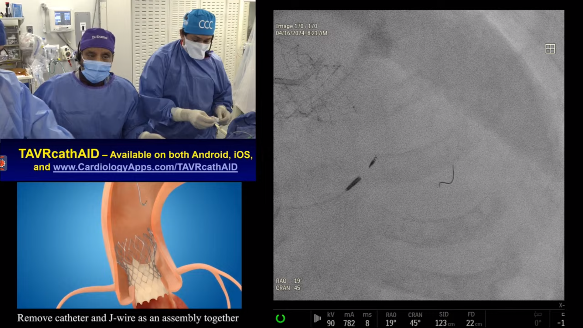
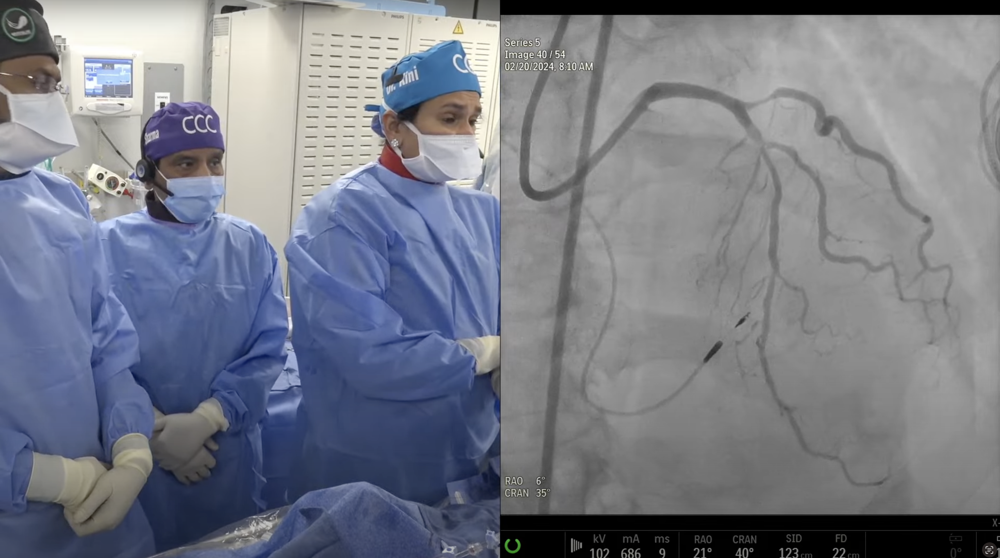
Case and Plan:
75-year-old female presented with new onset CCS Class II angina and positive stress MPI on November 9, 2020 revealing moderate apical and inferior ischemia. A Cardiac Cath on November 24, 2020 revealed 2 V CAD: 95% proximal RCA, angulated tortuous 95% mid LAD bifurcation lesion, LVEF = 60% and SYNTAX Score = 18. Patient underwent successful intervention of proximal RCA using Xience Sierra DES. Patient is now planned for staged PCI of extremely tortuous angulated mid LAD diagonal bifurcation lesion.
Q&A
Q
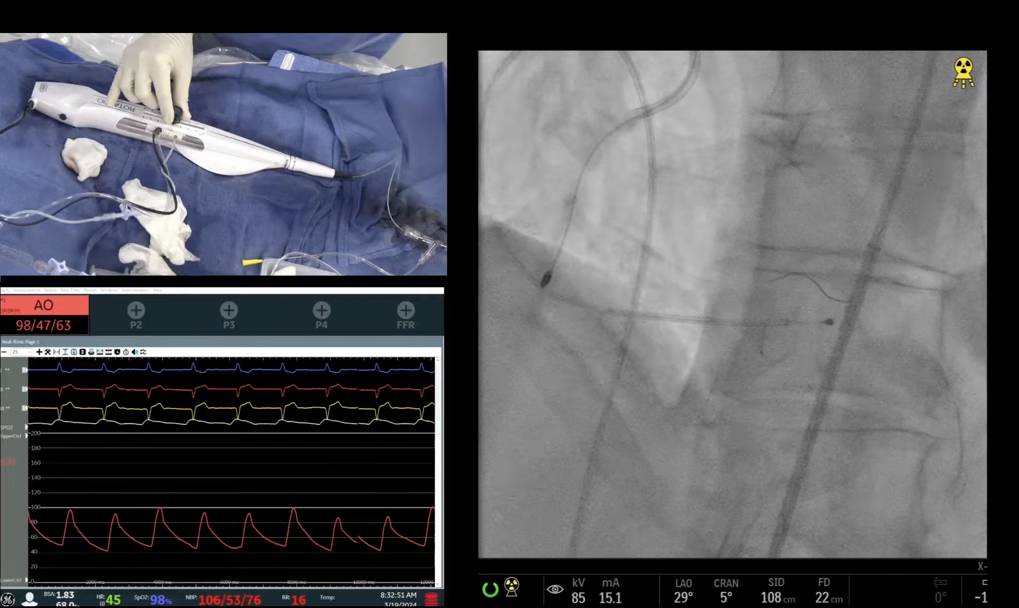
Had the SuperCross 120 not worked, what was your next plan of action?
A.
If SuperCross 120 did not work to cross the lesion, next step would have been stent across into the Diagonal and retry SuperCross or Venture catheter to cross into the distal LAD. Escalation to stiffer guide wires like MiracleBro 6-12, Confianza 12 or Progress 200T would have been tried before stenting across.
Q
If you had ended up using the Miracle wire, were you not concerned about this stiff wire causing a dissection plane?
A.
That is correct that many times, stiffer guide wires like MiracleBro or Confianza in angulated lesions can create the dissection plains, but it is the part of technique as long as guide wires re-enters into the distal lumen. Then balloon dilatation followed by stenting will create a new smooth working lumen. If guide wire can't get into the distal true lumen despite all efforts and wire tip escalation, then advise will be to end the procedure and bring back the pt for retry after 6-8 weeks. But if distal flow is compromised, then it needed to be treated accordingly with IABP, Impella or even urgent CABG depending on the pt's overall ischemic & hemodynamics condition.
Q
Have you any experience with the Crusade microcatheter?
A.
We have heard good reviews of the Crusade microcatheter but we have not used it in our cath lab. Fine cross followed by Corsair are our most common microcatheters.
Q
Which is your favorite deflectable tip catheter system?
A.
SuperCross 120 is our work horse deflectable catheter system followed by Venture OTW catheter.
Q
In this particular case, which bifurcating strategy would you have used for a two-stent approach?
A.
Because of large size side-branch Diagonal, minicrush 2-stent technique would have been our preferred 2-stent approach. If 6Fr guide catheter is used, then TAP or DK-crush techniques would have been other appropriate approaches.
Q
You have often advocated cutting balloons for ostial lesion modification prior to stenting? Did you consider it in this case?
A.
My personal experience (bias) of using cutting balloon in the ostial lesion is that it causes less dissection (if no stenting) and also results in better stent MLD. We did not use cutting balloon in today's case due to sharp angulation post stent placement and concerns of not crossing the lesion.
Q
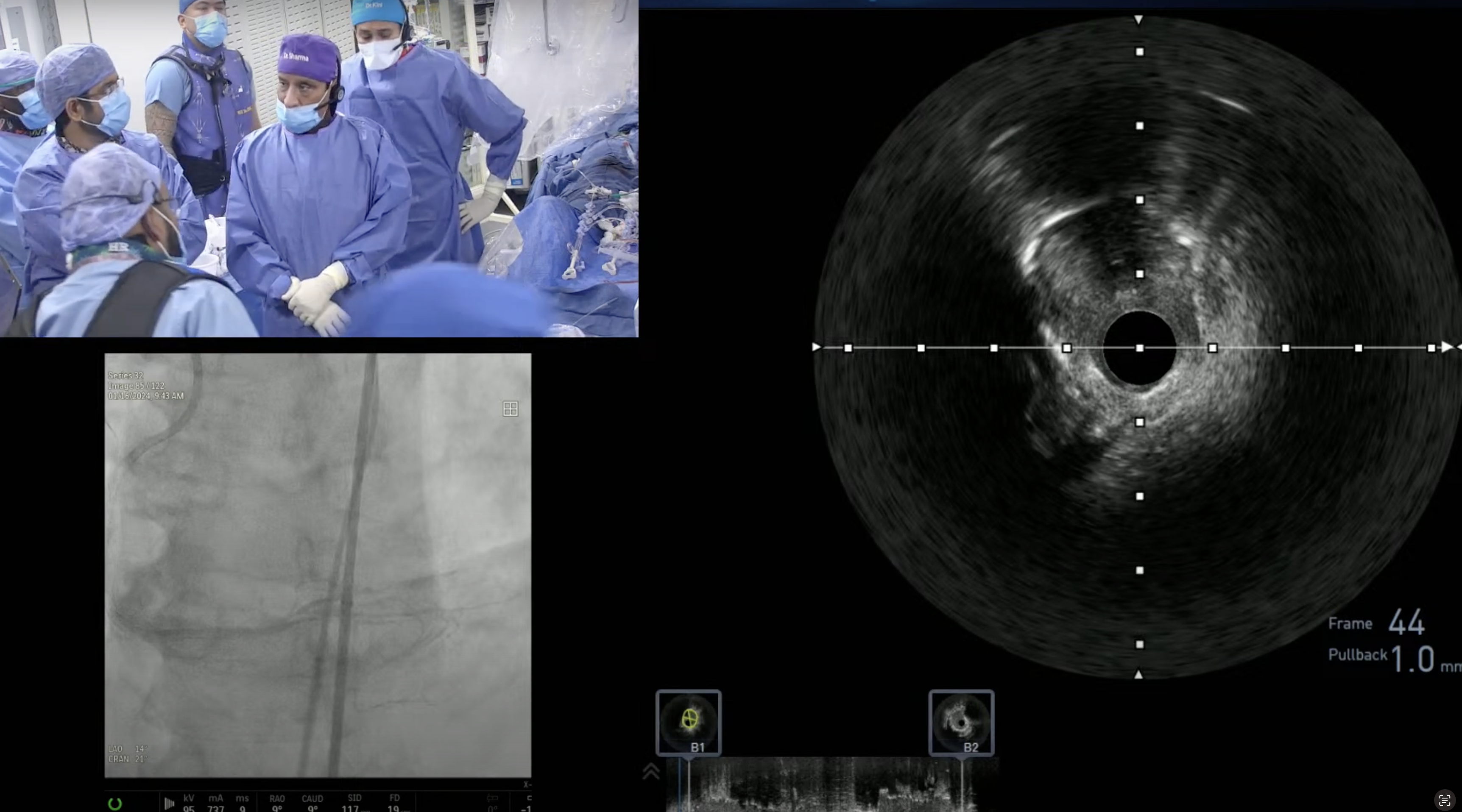
Does imaging help in selecting the microcatheter or guidewire approach for such cases?
A.
We don't usually use the intravascular imaging to select guide wire or microcatheters in these angulated cases. Imaging especially IVUS may help in selecting the point of entry into the side branch; also in making the appropriate curve on the distal guide wire tip.
Q
Which DES do you like for treating angulated lesions?
A.
All available DES are very conformable to the angulated lesions with minor variations in stent design and strut thickness. Xience or Synergy are our preferred stents in angulated lesions.
Q
What has been your experience with Asahi wires for such cases?
A.
ConfianzaPro 9 or 12 due to tapering tip of 0.010" helps to navigate thru to the distal lumen even after going sub-intimal for a short distance. Many times MiracleBro 6 due to its stiffer nature and 0.014" tip, will help in making the sharp turns in these angulated lesions.
Q
How do you deal with the situation of pseudo-lesions in angulated cases?
A.
Occurrence of pseudo-lesions in angulated case posses tough challenge but ballon dilatation followed by end to end lesion stenting will solve the problem with good angiographic results. Atherectomy (RA or OA) should be avoided in pseudo-lesions even if lesion is heavily calcified due to concerns of severe dissection and perforation.
Excellent case