CASE & Plan:
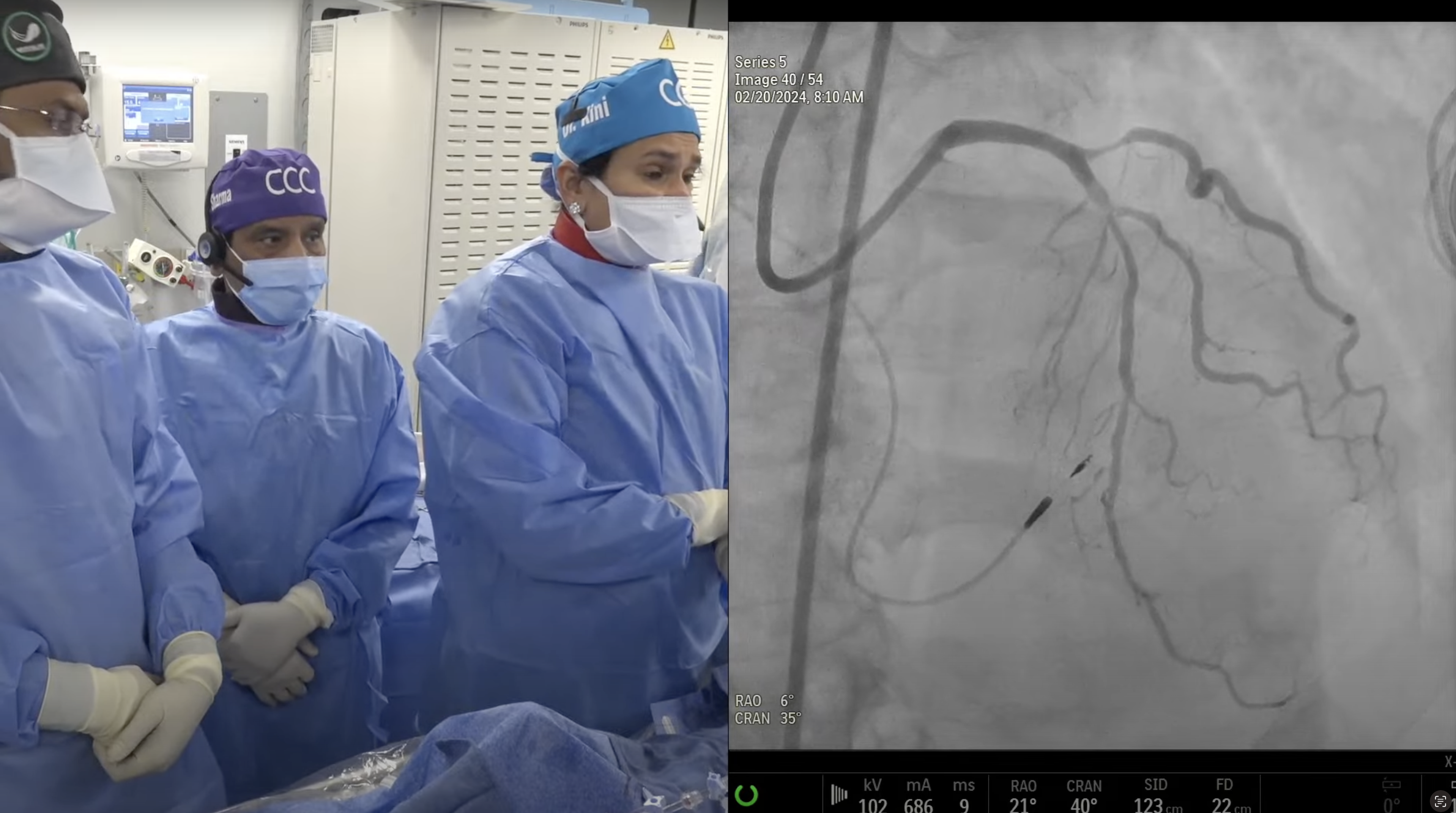
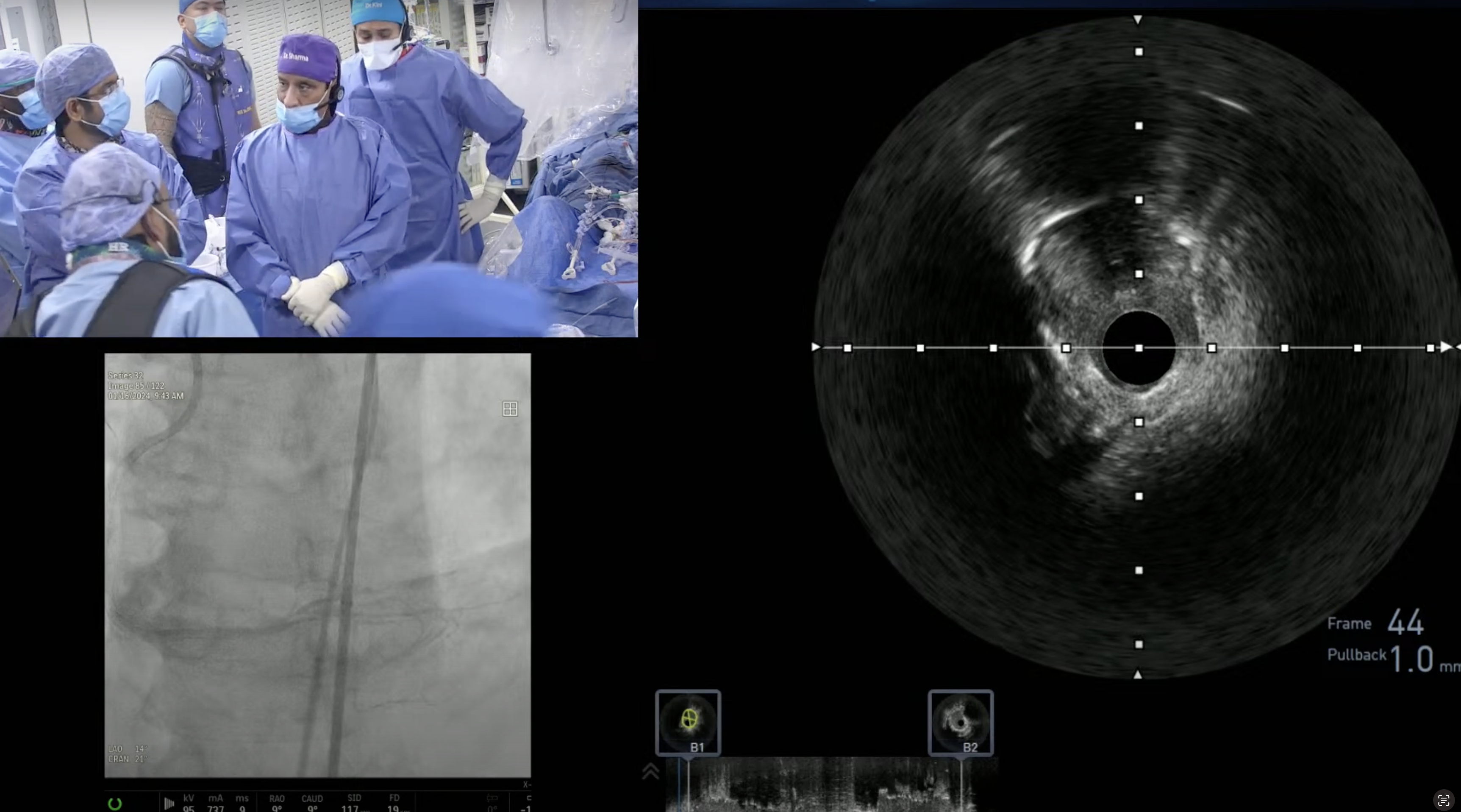
54-year-old male presented with CCS Class I angina and CTA revealed high Ca+ score and 2V CAD with +CT FFR. A Cardiac Cath on January 6, 2023 revealed 2 V CAD: 70% ostial and 80% mid LAD, 80% D1 bifurcation, subtotal LCx-OM2 with normal systolic LV function and SYNTAX Score of 25. Patient underwent successful intervention of LCx-OM2 using PTCA and Promus DES and did well. Patient is now planned for imaging guided staged PCI of LAD/D1 bifurcation using RotaTripsy and Mini-Crush stenting.
Q&A
Q
What would have prompted you to use IVL in this case?
A.
If NC balloon after Rota does not fully expand, then IVL use is appropriate before stenting. In the past we use to upsize the burr in this scenario (8-10% of cases) which we now rarely do (1-2% only). Hence we just use IVL as adjunct after Rota in large vessels which almost always works.
Q
Could you have used the same Rota burr for the ostial of the diagonal branch instead of using the cutting balloon?
A.
Diagonals, due to their angulation are notorious for dissection and perforation with Rota, hence should be avoided as much as possible. That is why we recommend CB in moderate+ calcified Diagonals. IVL is rarely needed in this situation.
Q
After using the balloon for the ostium of the LCX, why leave this critical area with a higher possibility of restenosis?
A.
There are 2 ways to look into this issue of LCx ostial PCI, stent or not stent after crossover stenting. Our strategy is that if there was <30% lesion pre-stenting, and ostial pinch occurs after stenting (most likely from the plaque shift), then try not to put the bail out stent and come out with only final KBI using low pressure balloon inflation in LCx ostium. Yes, if ostial LCX still remains 70%+ narrow or dissected after KBI, then bailout stenting with TAP (preferred) or reverse crush will need to be done; in our case it was plaque shift and residual ostial LCx lesion after KBI was about 50-60% without dissection and hence was not stented.
Q
Could this LCX ostia be stented?
A.
Yes as I said earlier, LCx ostium can be stented (if remained severe angiographically after KBI) using TAP, reverse crush or T stenting techniques; all followed by final KBI.
Q
What are your present criteria for RotaTripsy?
A.
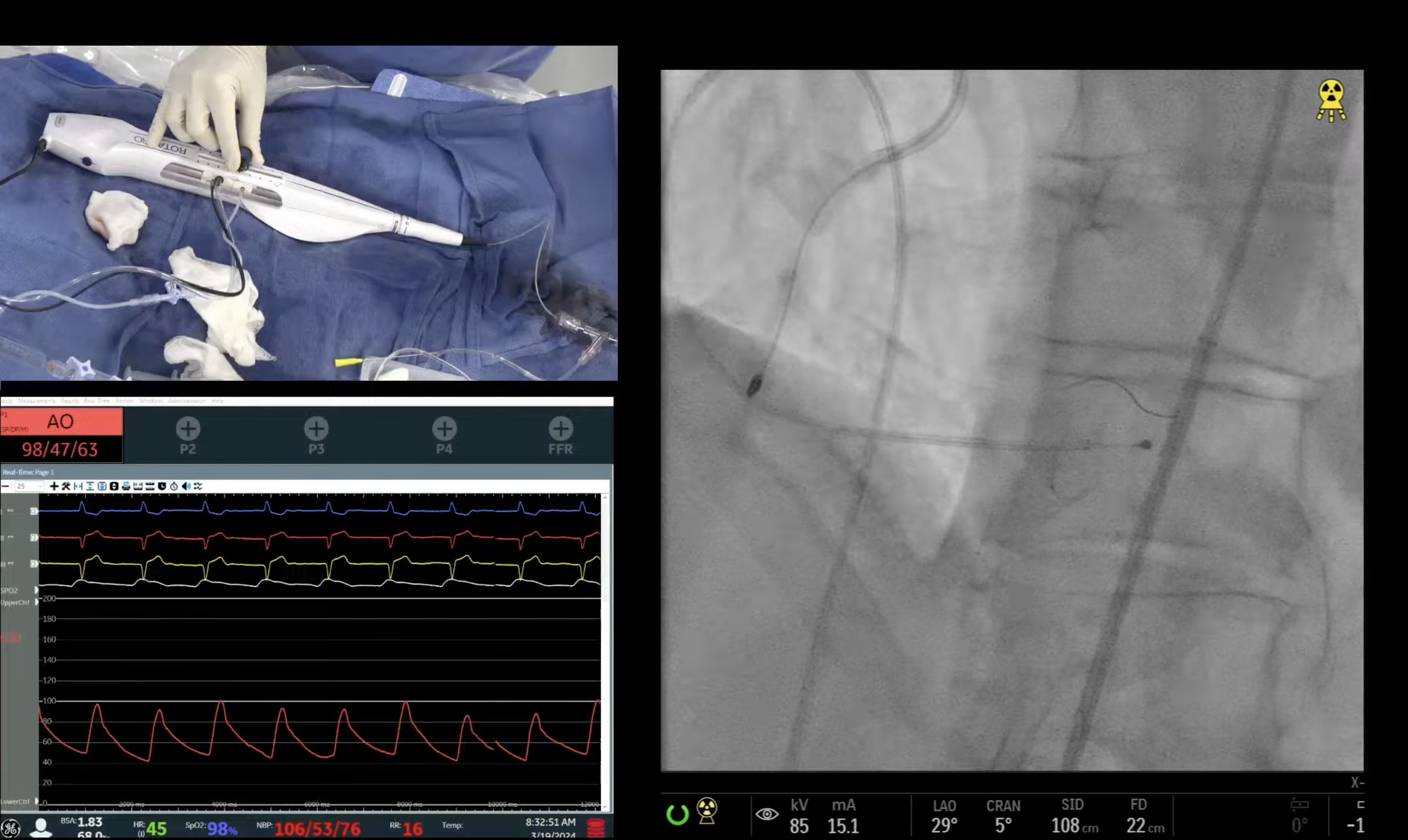
Severely calcified significant (>90%) lesion in a large vessel (>3.5mm) with concentric Ca++ or calcific nodule are the ideal case for RotaTripsy by using 1.5-1.75mm Rota burr followed by 1:1 size IVL balloon dilatation.
Q
Any new developments with Orbital Atherectomy (OA)?
A.
Last new device development of OA are; 1) Glide assist allowing crown to be advanced easily to distal lesions and easy device removal; 2) Viper Wire Flex tip which is hydrophilic wire with better torqueability and minimize wire bias in tortuous lesions.
Q
Based upon your superb presentations from your institute on antiplatelets, do you see these prompting change in guidelines?
A.
Three recent publications from our institution highlighted 3 important messages; 1) Deescalation from Ticagrelor or Prasugrel to Clopidogrel is safe and preferred after 1-3 mths of PCI; 2) All 3 antiplatelet agents are safe and effective once a clinical decision is made considering clinical and angiographic lesion features even in stable CAD; 3) There is no difference in death or MI at one year with use of Ticagrelor vs Prasugrel in contemporary clinical practice is all comers (Stable CAD and ACS) This is in contrast to ISAR-REACT 5 trial which showed superiority of Prasugrel over Ticagrlor in ACS pts.
Q
What are your present recommendations for Clopidogrel?
A.
Still 60% of all PCIs at MSH receives clopidogrel especially elderly and high bleeding risk pts.
Q
And for Prasugrel and Ticagrelor?
A.
Either of them (Tic or Pra) preferred in ACS especially STEMI and complex CAD in a pt with low bleeding risk and in young Pt’s.
Q
When and how are you using PRU assays?
A.
We are now rarely using PRU assay (value of 230U 2) High risk PCI where clopidogrel is indicated clinically, 3) Any pt with Stent Thrombosis or recurrent restenosis on Clopidogrel.


