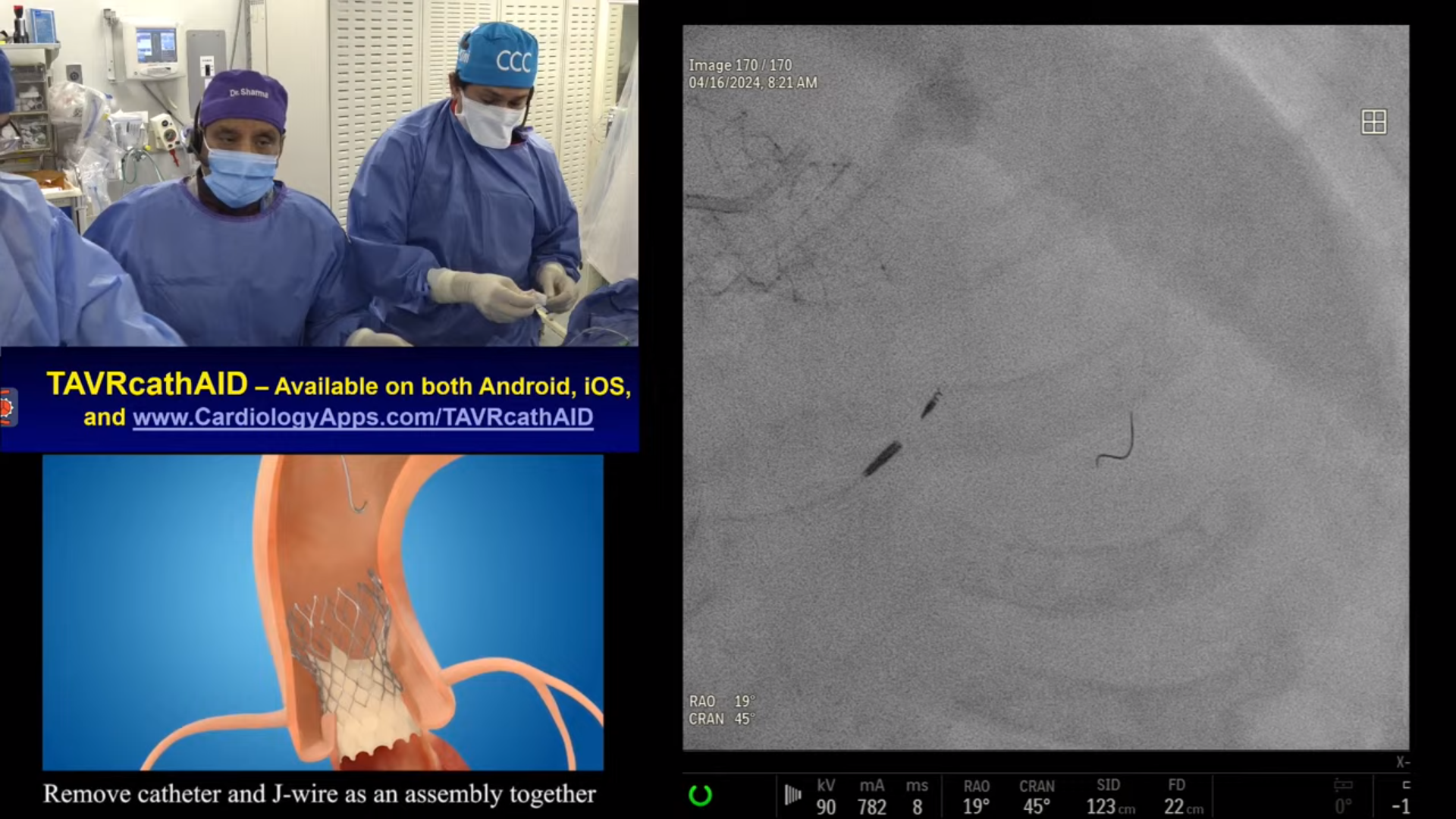
72 year-old male with strong F/H of CAD and mild dyspnea on moderate exertion, had high risk stress echo for anterior wall ischemia and mild AS (AVA 1.3 cm2 and mean gradient = 20 mmHg). A Cardiac Cath on June 10, 2016 revealed 2V CAD; 70% calcified proximal LAD with FFR of 0.76, 80% D1 with FFR of 0.81, 70% LPL with FFR of 0.87. Anti-ischemic therapy was started. Patient is now scheduled for Rotablator + PCI of LAD and Diagonal Lesions using Absorb GT1 with OCT guidance.
Moderator: Sameer Mehta, MD
Q&A
Q
What BVS engineering changes should be sought?
A.
Most important being decreasing the strut thickness yet maintaining the tensile strength. Also making it radio opaque.
Q
Plaque modification and lesion preparation in every BVS case?
A.
Lesion preparation in all cases by predilatation of the lesion with NC balloon and plaque modification using Cutting ballon in moderately calcified lesion and RA or OA in severely calcified lesions, will be essential before BVS implantation.
Q
Should imaging be mandatory or recommended?
A.
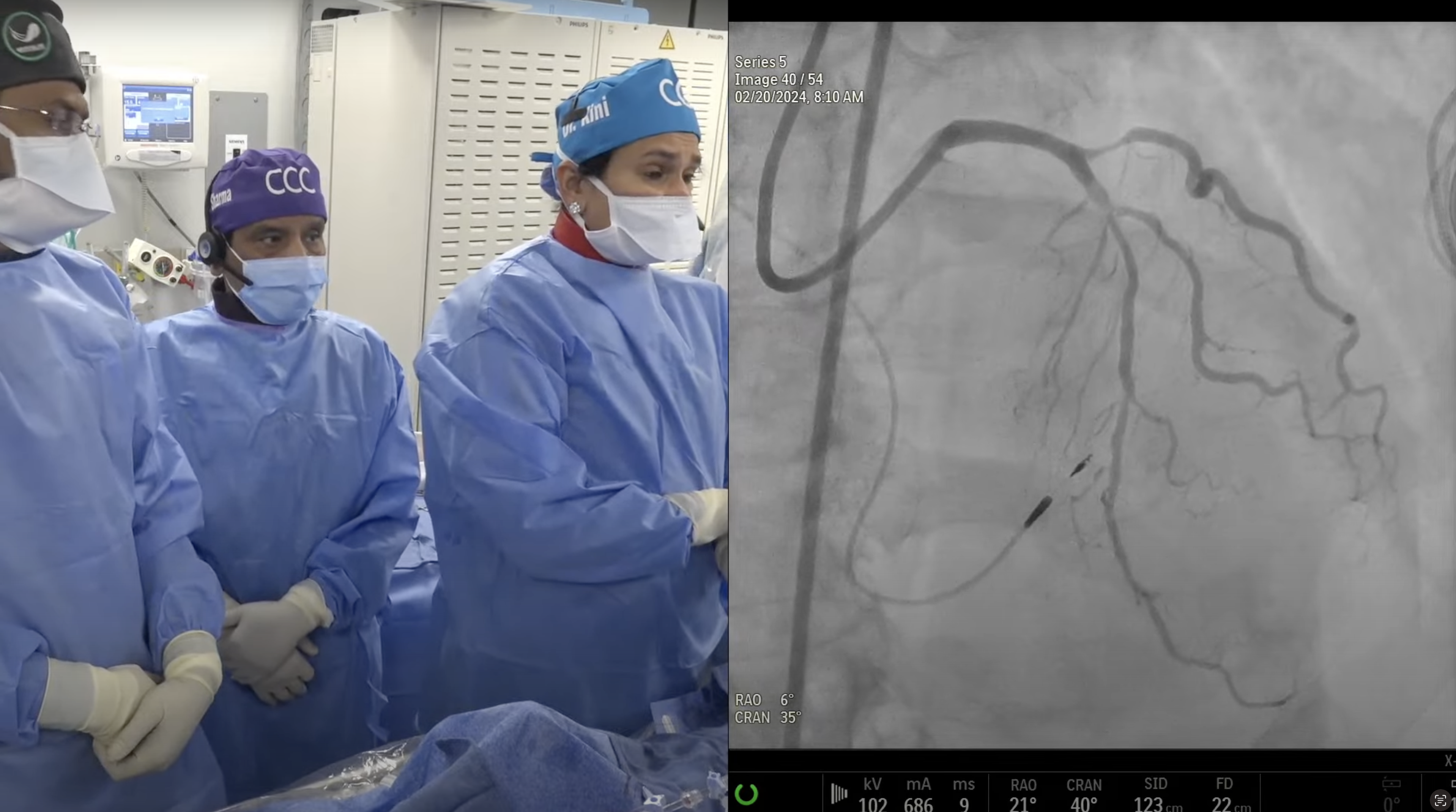
I will recommend imaging preferably OCT, being essential in first 15-20 cases to exactly understand the BVS deployment characteristics. All potential small vessels of 2.5 mm size should have imaging to confirm the vessel size of >2.5mm as vessels <2.5mm have bad outcome after BVS.
Q
Greater than 0.5 mm post expansion - detrimental to struts?
A.
Experimental data have shown scaffold damage once balloon size was 1.00 mm higher then the recommended size. Hence upto 0.5mm post dilatation is recommended. But if still there is a 10-20% residual stenosis after 0.5mm higher size post-dilatation, then going to 0.75mm oversize will be ok rather then leaving the less then optimally dilated scaffold.
Q
What are particular tips for overcoming lack of visibility?
A.
Good X-ray equipment and paying attention to the radio-opaque markers at both ends of BVS. Stent boost is also helpful.
Q
What should be post expansion strategies for BVS in bifurcating lesions?
A.
In bifurcation lesions, avoid the kissing balloon technique and just open the SB by a small size ballon at 6-8 atm.
Q
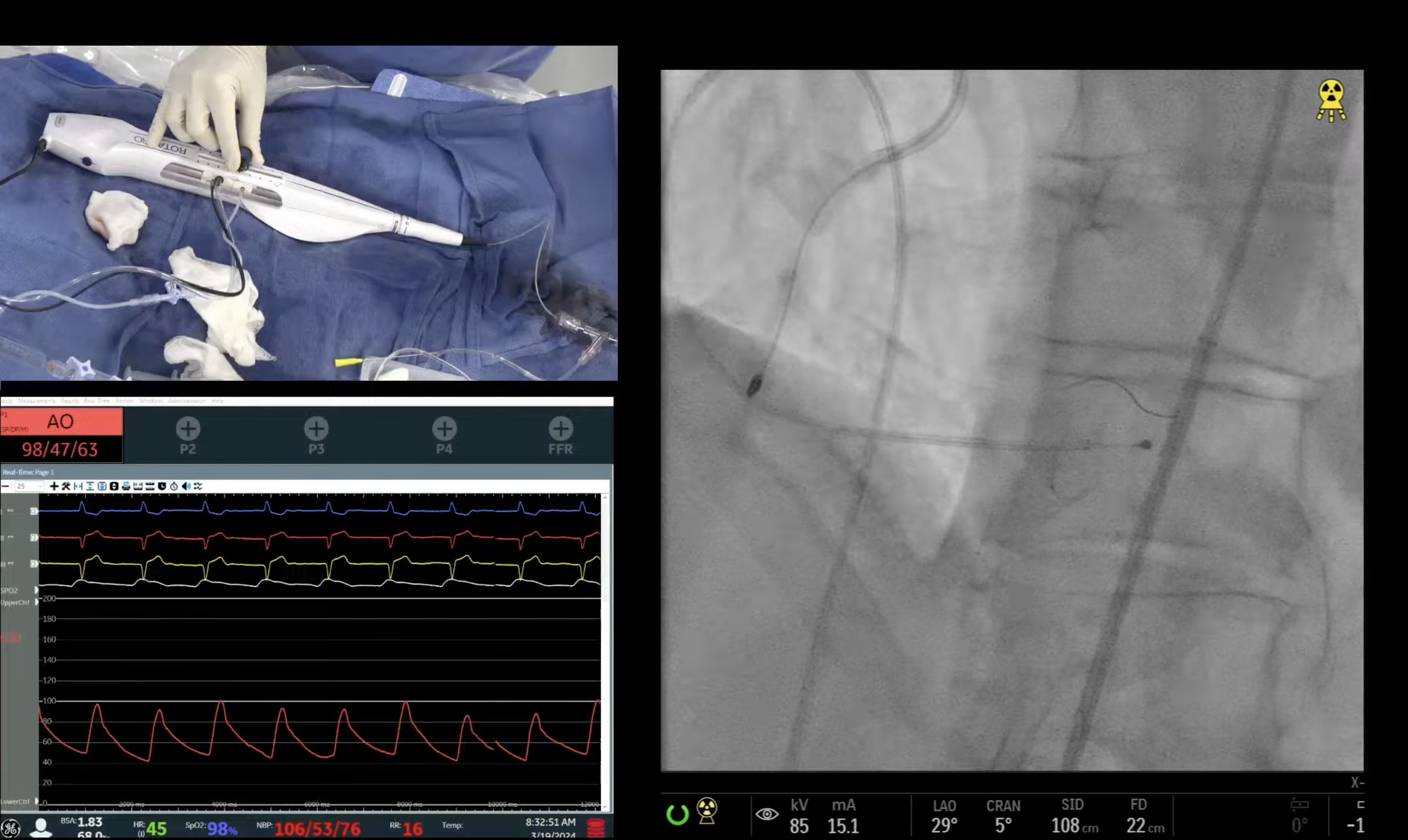
Could your case for today have been done by other plaque modification techniques besides Rotablator?
A.
Since lesion was severely calcified and hence atherectomy (laser, rotational or orbital) is essential before BVS deployment.
Q
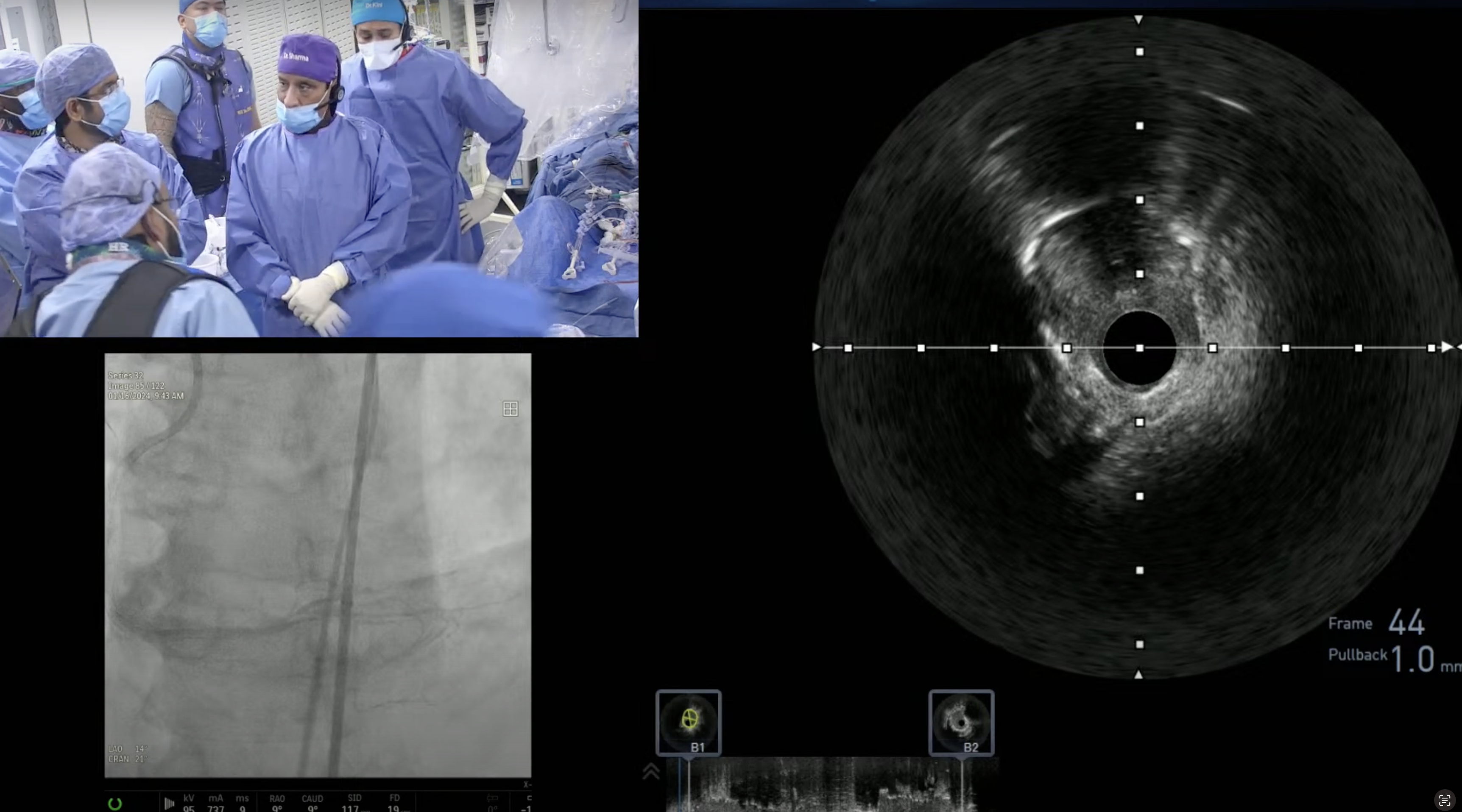
What is more accurate for sizing - IVUS or OCT?
A.
In my option IVUS is better for vessel sizing while OCT is better to evaluate BVS expansion and residual dissection.
Q
Is most scaffold thrombosis technique related?
A.
I will say by enlarge, scaffold thrombosis is mainly technique related (2/3rd) along with DAPT noncompliance responsible in 1/3rd.
Q
What are the three best techniques to avoid scaffold thrombosis?
A.
Three best techniques to avoid scaffold thrombosis are;1) avoid BVS in vessel size <2.5mn, 2) routine lesion preparation and avoid BVS if lesion could not be fully expanded and 3) routine post dilatation with an appropriate size NC balloon at 18-22 am.