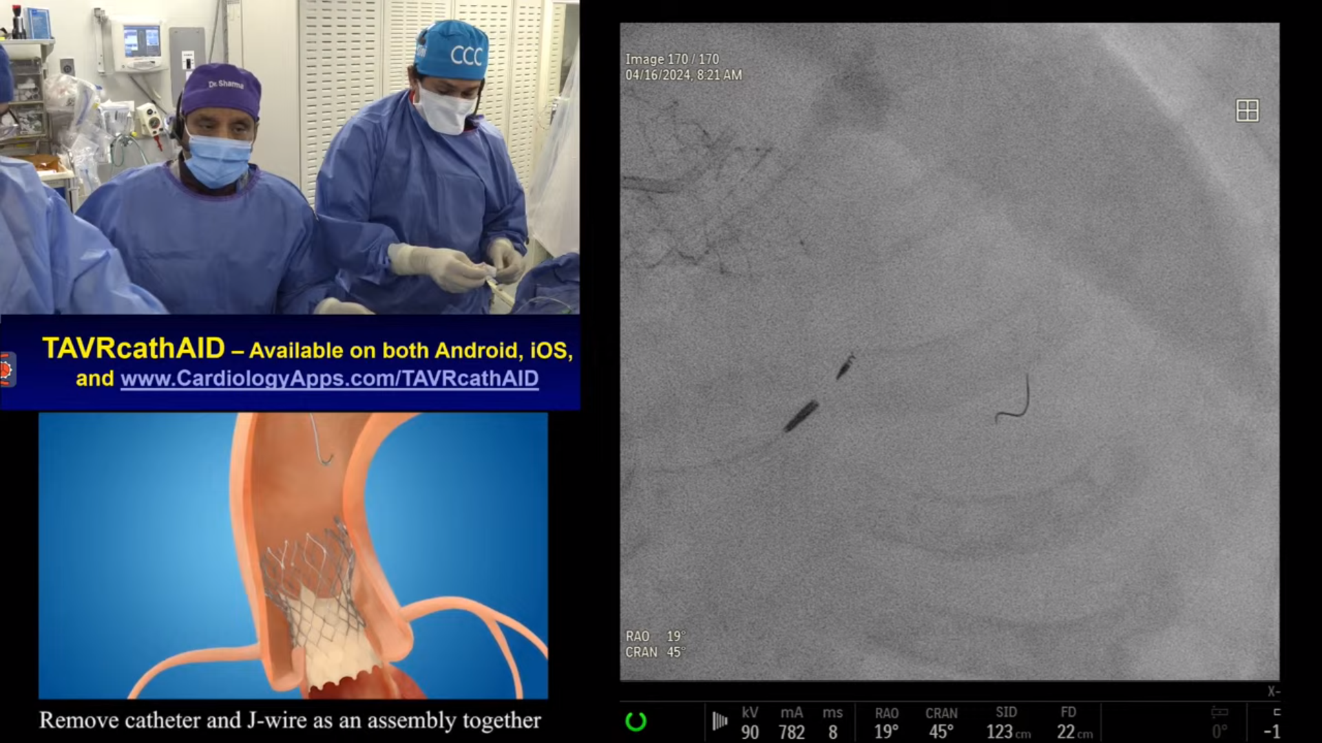
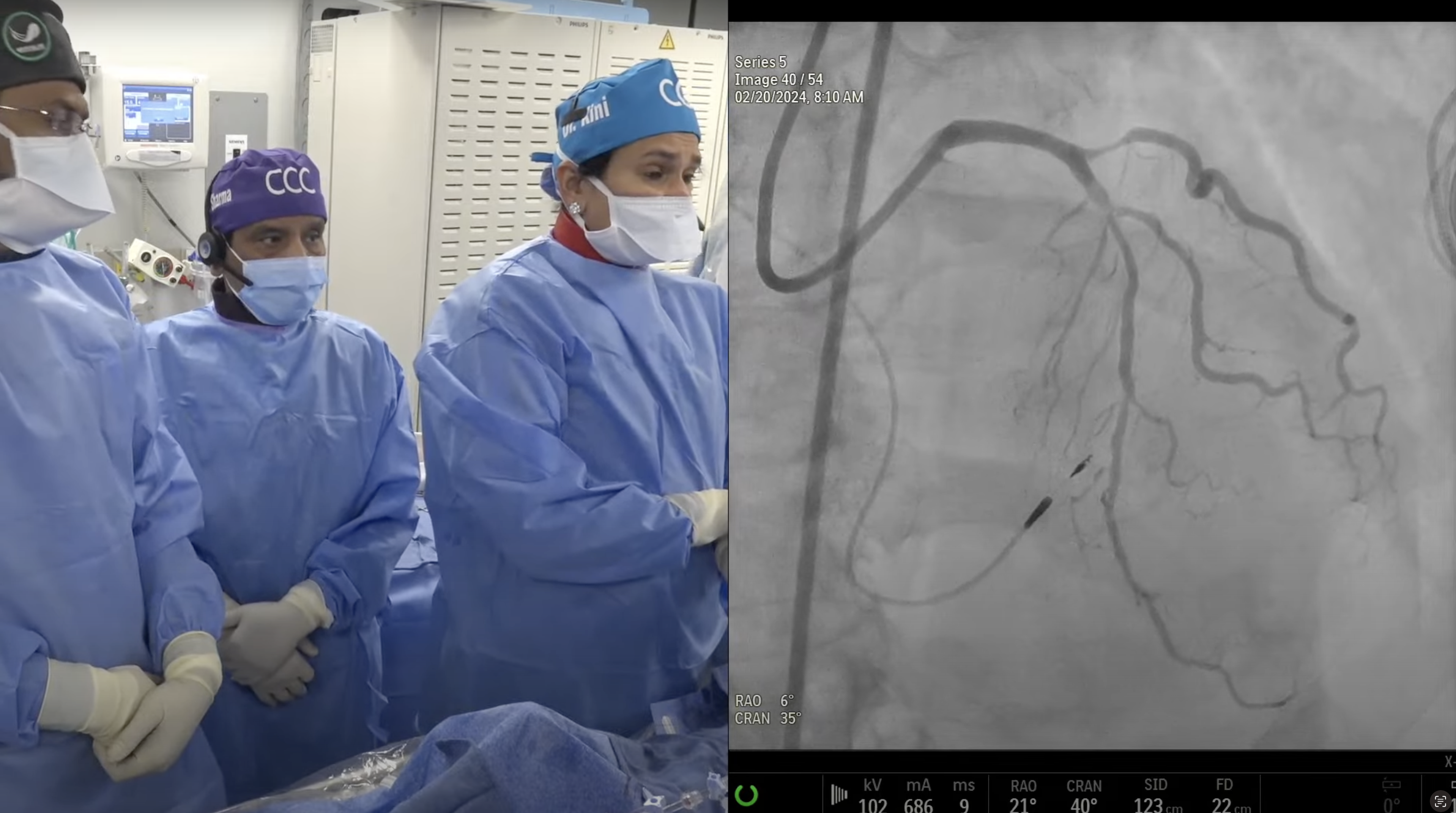
83 year-old female with new onset crescendo CCS Class III angina and dyspnea with known moderate left main disease since 2009. A Cardiac Cath on January 12, 2015 revealed calcified distal left main bifurcation: 80% distal LM, 70% proximal LAD and 90% proximal LCx with normal systolic LV function; SYNTAX Score 24. Heart Team discussion took place and patient elected for PCI. Patient is now planned for PCI of calcified dLM bifurcation using OCT, rotational atherectomy and planned dedicated two stents approach.
Q&A
Q
What were the major challenges formulating your very effective Appropriateness Criteria at your hospital for which you are increasingly being viewed as a leader?
A.
Most important challenge was convincing the referring physicians to start maximal medical therapy first once pt presents with angina symptoms or mild-mod ischemia. Referral for Cath should then be second step for continued symptoms.
Q
Is there a specific committee that was created?
A.
It was just repeated education of the physicians first and then the nurses and NP who reviewed the precath note and noninvasive tests day before. If felt inappropriate to Cath bases on the information available, then I took the responsibility first calling the referring physician and then pt about rescheduling the Cath procedure at a later date after starting the MMT.
Q
Is a cardiac surgeon involved?
A.
Cardiac surgeon is not involved in implementing the AUC criteria of PCI. Yes they are involved as the Heart team discussion in the complex PCI of high Syntax score, to make the final decision about revascularization; CABG or PCI.
Q
In effectively monitoring this policy, is it challenging to deal with referring physicians?
A.
Yes it was difficult in the beginning to explain to referring physician that why PCI was not done despite a 80-90% lesion. But with education and providing the rationale of deferring PCI, all were satisfied. Later routine use of FFR and deferring PCI if FFR was >.8, really helped us to satisfactorily convincing the referring physician.
Q
Do your speak about these issues nationally and internationally?
A.
Yes as every ACC meeting for last 3 years, I gave the talk or debated the AUC use and it's utility as a quality parameter. We had one theHeart.org sponsored program in 2012 on this topic.
Q
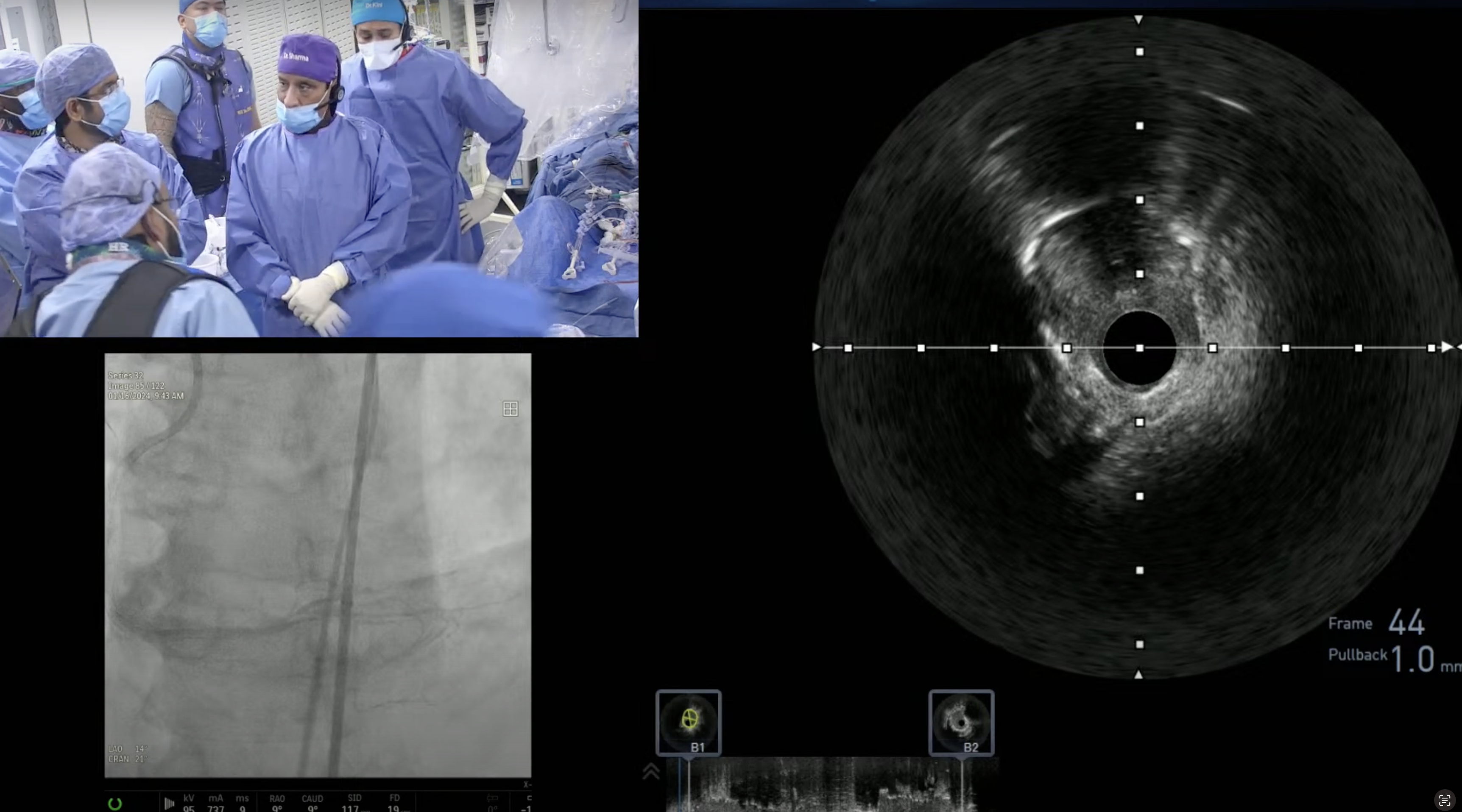
How does OCT compare to IVUS for assessing calcification?
A.
Both are good techniques but I personally believe that IVUS is superior to OCT in really quantifying the extent and degree of calcium in the lesion.
Q
If there is reluctance to use Rotablator, which is more effective for debulking - Cutting Balloon or Angiosculpt?
A.
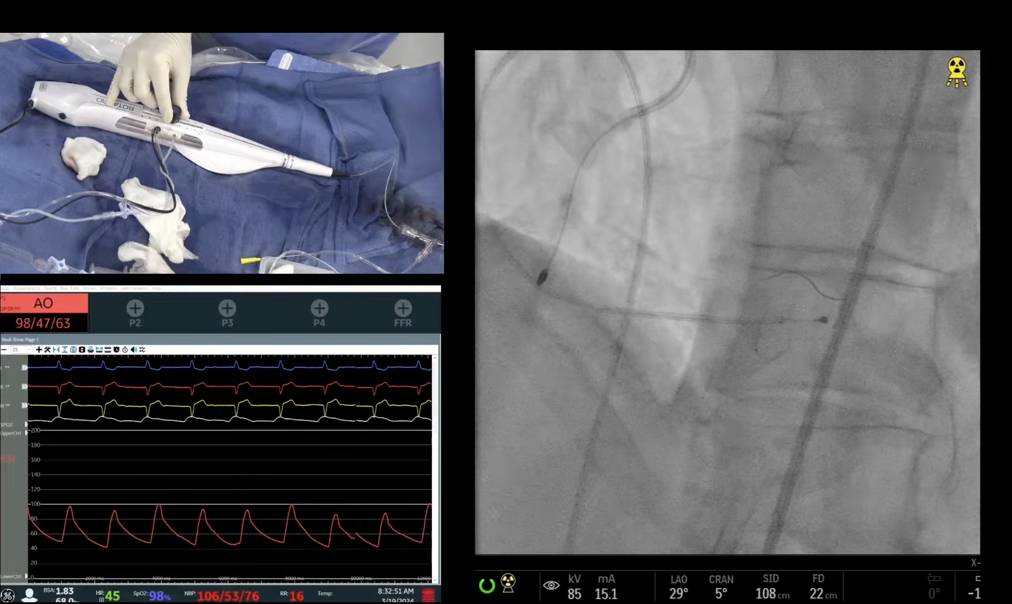
Yes Atherotomy techniques of Flextome and AngioSculpt can be tried in moderate-severely calcified lesions. But in heavily calcified lesion with tram track calcium, Rotational or Orbital Atherectomy use is must; as lesion will not be crossed or cracked otherwise.
Q
What caused the proximal LMCA dissection - guiding catheter related injury or it was simply from the complex maneuvers for a very difficult procedure?
A.
Yes Type B intimal dissection in prox LM in our case was likely due to 2 oversized stents with proximal balloon edge. We then watched the pt for 15 minutes and a f/u Angio shot showed no progression of dissection and hence was left alone. Otherwise it would have required 2 additional stents in each SKS limb proximally to cover the dissection. Another way in these dissection cases will be to put one DES proximally to cover the dissection after crushing the sidebranch stent by PTCA followed by final kissing balloon dilatation.
Q
What strategies are effective to reduce bleeding in an elderly female patient?
A.
Most important strategy to minimize the vascular bleeding is proper vascular access, use of vascular closer devices and avoid GPI use beyond 1-2 Integrilin blouses. PCI with Bivalirudin use has consistently shown to reduce both access as well as non-access site bleeding.
Q
No role for surveillance angiography?
A.
2010 LM PCI guidelines, have eliminated routine surveillance angiography at 3-4 mths post ULM PCI to detect any early restenosis. I still recommend follow up angiography in about 20-25% of total ULM PCI cases, especially in pts with 2+ stent use and pts with residual lesions left.